DIRS
DIRS

Diffuse Raman Spectroscopy (DIRS)
Diffuse Raman Spectroscopy (DIRS) combines the high chemical specificity of spontaneous Raman scattering with the aptness of Diffuse Optics to investigate highly diffusive media non-invasively. This field has witnessed an impressive growth due to the introduction of Spatially Offset Raman Spectroscopy (SORS) which permits to probe diffusive media in-depth, leading to flourishing applications in the field of clinical diagnostics, pharmaceuticals, cultural heritage, safety and security. Our DIRS laboratory exploits a novel approach based on time-domain detection of diffuse Raman photons migrating through the scattering material, permitting depth-resolved reconstruction of Raman emitters based on the photon traveling time.
Development of advanced techniques for Diffuse Raman Spectroscopy
(referent prof. Antonio Pifferi)
The DIRS laboratory has explored novel approaches to extract spontaneous Raman signals in-depth from highly diffusive media. Three techniques have been proposed so far:
- Frequency Offset Raman Spectroscopy (FORS): Raman signals emitted at different depths within the scattering medium can be discriminated using multiple excitation frequencies. The proposed methodology obtains depth sensitivity exploiting changes in optical properties (absorption and scattering) with excitation wavelengths. The approach was demonstrated experimentally on a two-layer tissue phantom and compared with the already consolidated spatially offset Raman spectroscopy (SORS) technique. FORS attains a similar enhancement of signal from deep layers as SORS, namely 2.8 against 2.6, while the combined hybrid FORS-SORS approach leads to a markedly higher 6.0 enhancement.
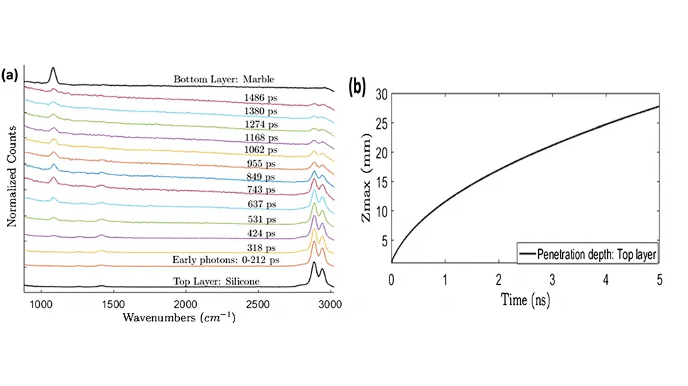
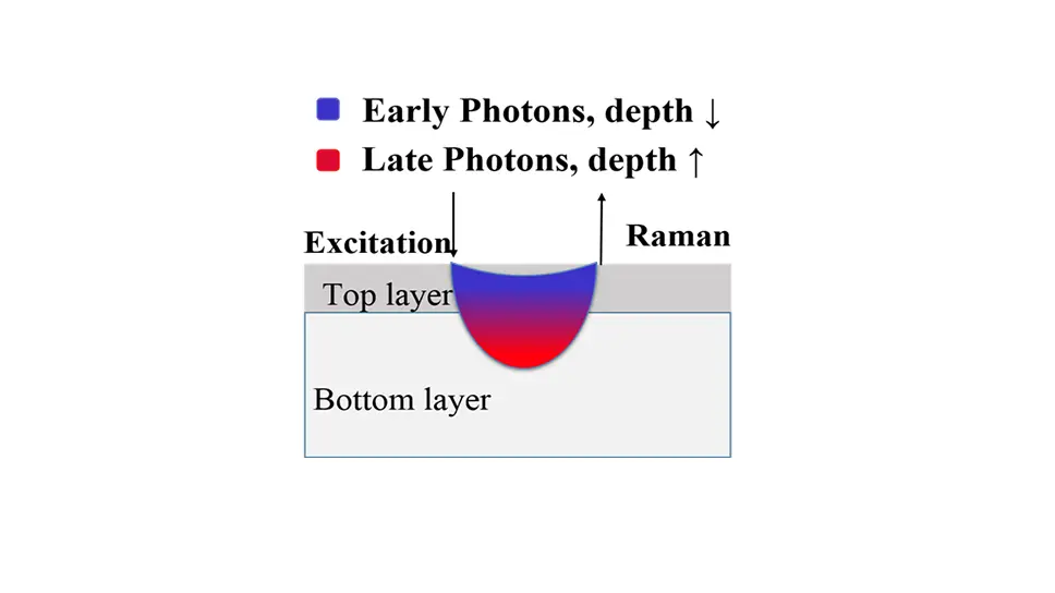
- A different approach is based on a novel time-correlated single-photon counting (TCSPC) camera that simultaneously acquires both spectral and temporal information of Raman photons. Depth sensitivity is achieved by time gating Raman photons at different delays. Time domain can provide time-dependent depth sensitivity leading to a high contrast between two layers of Raman signals.
- A third approach exploits compress sensing to use a single time-resolved single-photon detector for temporal and spectral acquisition of diffuse Raman signal. A Digital Micromirror Device (DMD) inserted within a Raman spectrometer modulates the spectral signal detected by a single-pixel time-resolved detector. The adoption of a proper orthogonal basis (e.g. Hadamard) and the corresponding inverse transform permits to reconstruct the Raman signal both temporally and spectrally.
Figure 2 – Diffuse Raman Spectra as a function of photon arrival time (a). Maximum depth reached by photons as a function of photon travelling time (b), from Ref.2.
Development of theoretical models and reconstruction strategies for Diffuse Raman Spectroscopy
(referent prof. Antonio Pifferi)
Theoretical models stem from the Diffusion Approximation of the Radiative Transport Equation were derived to describe Raman emission and propagation through highly diffusive media in the time-domain.
These models were used for the reconstruction of Raman signals from layered media exploiting the photon arrival time to disentangle signals generated at different depths. The following step is the development of full 3D Diffuse Raman Tomographies.
Equipment
- Tuneable active mode-locked Ti:Sapphire laser for narrowband tunable raman excitation in the 680-1100 nm range with <50ps temporal resolution
- Time-resolved, single-photon raman spectrometer based on compress sensing single-pixel detection
- Non-contact Raman probe for diffusive media with ring illumination and variable source-detector distance


Research projects
Fast gated superconducting nanowire camera for multi-functional optical tomograph (fastMOT)
Find out more