Development and application of innovative instrumentation for breast imaging and spectroscopy
Development and application of innovative instrumentation for breast imaging and spectroscopy
Breast cancer is the most common cancer and one of the main causes of death in women in Europe and the United States, as well as in many developing countries.
Optical diagnostic methods are intrinsically non-invasive and could therefore be effectively used both for screening and monitoring, when repeated measures over time are required. By estimating the optical properties (absorption and scattering), it is possible to derive the composition of tissue and some information on its microscopic structure. Furthermore, functional information can be obtained. Finally, the instrumentation is generally quite simple and inexpensive, compared to that currently used by many medical diagnosis techniques.
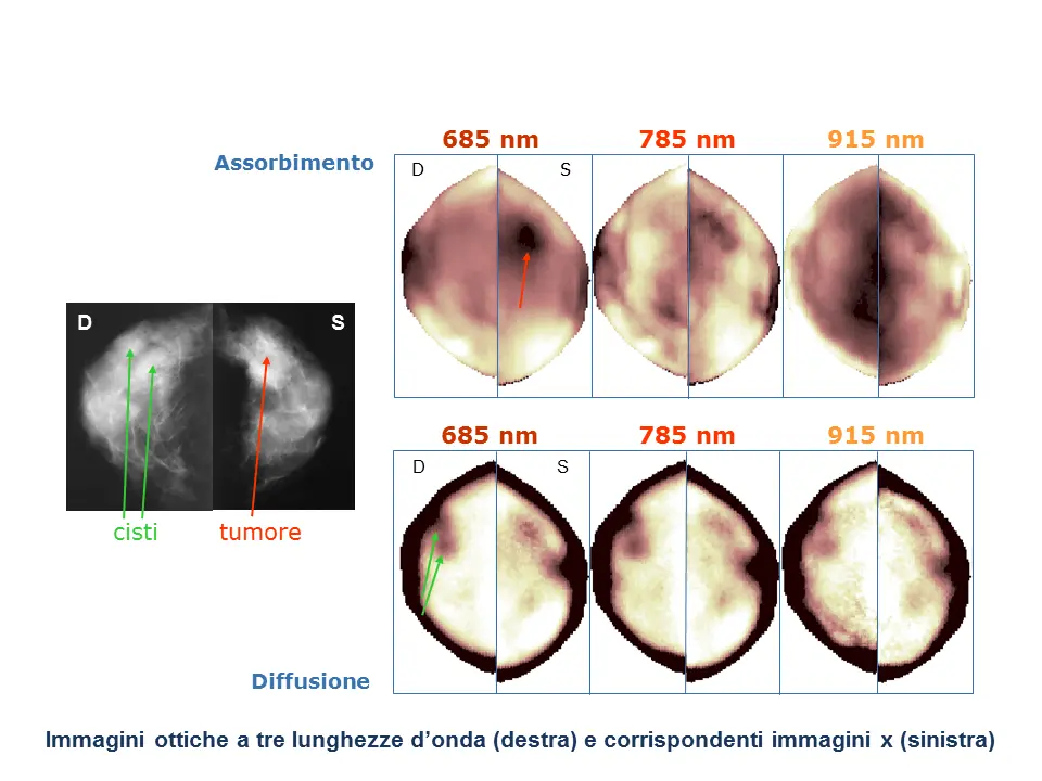
The approach chosen by our research group at the Department of Physics involves the evaluation of the optical properties of tissue by time domain transmittance measurements performed at several wavelengths in the red and near-infrared. A short pulse of light (~ 100 ps) is injected into the tissue. The transmitted pulse is detected and, (typically) using the diffusion theory, the average value of the absorption and scattering coefficients of tissue is obtained. By repeating the measurement at multiple wavelengths, it is possible to evaluate the absorption of the different constituents of tissue and retrieve its composition. From the knowledge of the scattering properties instead we obtain information on the microscopic structure of tissue.
Master's Degree theses are available for two diagnostic applications with distinct aim and implementation. In both cases, the activity involves several aspects: from the design and development of a tool for clinical use to the characterization of its performance, up to the interpretation of optical data for diagnostic purposes and to the actual clinical application. The two activities are described briefly below.
Estimate of cancer risk related to breast density
Breast density (i.e. the fraction of fibroglandular tissue in the breast) is recognized as an important risk factor for the development of tumors. Currently breast density is estimated by analyzing x-ray mammograms. A non-invasive method of evaluating tissue composition, not requiring the use of ionizing radiation, such as the optical one, would allow an extensive application, even on young women, allowing the identification of high risk subjects, which could then follow a personalized screening and/or diagnostic path, for early diagnosis and preventive interventions aimed at reducing the risk itself (e.g. by acting on lifestyle).
Moreover, the technique developed at the Department of Physics, unique at international level, allows the non-invasive measurement of collagen content in tissue. Collagen is investigated because it contributes to tissue density, but also because it is involved in cancer development and progerssion. The estimate of collagen concentration in tissue could therefore have diagnostic relevance also as a risk factor independent of density.
We plan to develop and apply a tool for the evaluation of the average tissue composition by acquiring time-resolved transmittance data over a wide spectral range (600-1100 nm). Different acquisition geometries will also be evaluated (in particular reflectance measurements at various distances). The tool will be developed using innovative detectors and acquisition techniques.
Monitoring of neoadjuvant chemotherapy
For the treatment of breast tumors the use of neoadjuvant chemotherapy is increasingly widespread, aiming at reducing tumor size before surgery, for a more conservative intervention or, in the best cases, to completely eliminate the tumor.
Almost one in two patients does not respond to neoadjuvant chemotherapy, with high morbidity for the patient and high costs for the healthcare system. No non-invasive modality is currently available to monitor efficacy or predict therapeutic response. However, there is initial encouraging experimental evidence that optical techniques could effectively perform this function.
We have developed an optical mammography tool that allows to acquire maps of the average tissue composition by continuous acquisition of transmittance data resolved in time at 7 wavelengths in the red and near infrared (600-1100 nm). The instrument presents significant innovations at a technical level, which aim to obtain high quality measurements in times compatible with a clinical measurement. His clinical validation has just begun. The activity will now focus on the acquisition of clinical data and on the development and application of methods and tools for the analysis of clinical data.