OMT
OMT
Optical Projection Tomography is a three dimensional imaging technique which is particularly suitable to study millimeter sized biological samples and organisms. Similarly to x-ray computed tomography, OPT is based on the acquisition of a sequence of optical transmission (or fluorescence) images of the sample at several orientations. The acquired images, or projections, are combined to reconstruct the 3D volume of the sample, typically using a back-projection algorithm.
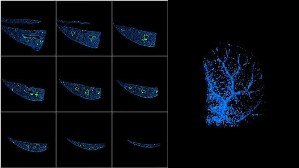
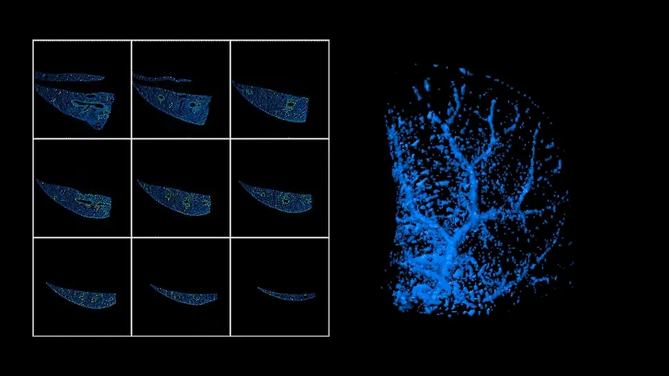
We develop OPT systems for a variety of biological applications, including oncology and spinal cord injury and neuroregeneration. Here is an example of a murine lung visualized with one of our OPT systems. Transverse sections of a healty lung, obtained measuring the autofluorescence of the tissue are shown on the left hand side and a 3D rendering is shown on the right hand side. The bronchial tubes are visible in both images. (fig.1)
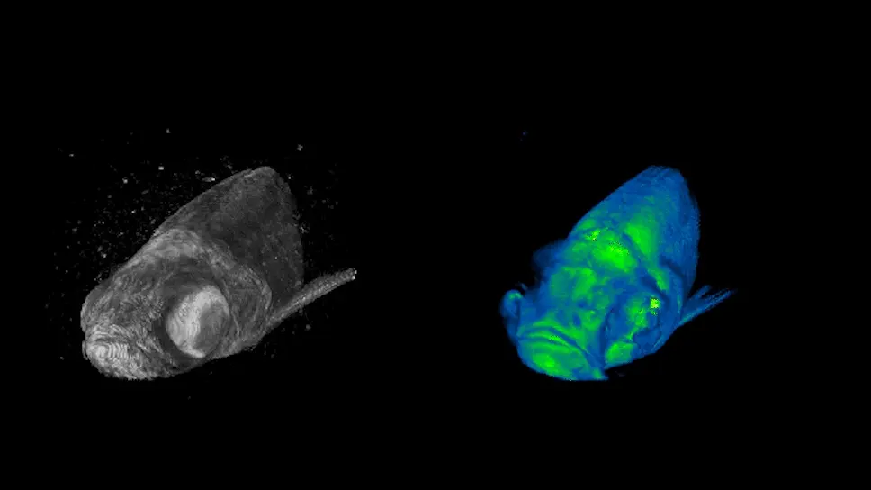
Recently we focused on the study of zebrafish (Danio rerio), a model organism widely used in developmental biology. Here are some examples of OPT scan on adult zebrafish, obtained by measuring the transmitted light (left) and the autofluorescence of the sample (right) (fig.3).
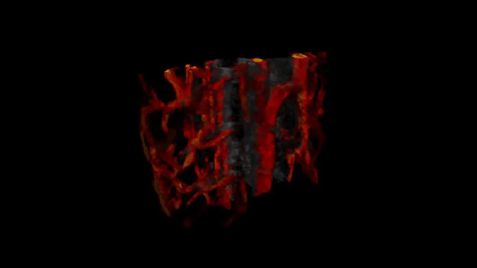
OPT can be operated in-vivo on juvenile zebrafish and embryos. We demonstrated that it is possible to image in 3D the vascular network of a zebrafish by detecting the movement of the cells in the bloodstream, without the injection of a fluorescent probe or the use of fluorescent reporters.
Here is an in-vivo label-free 3D reconstruction of the vasculature of a juvenile zebrafish trunk (fig.2).
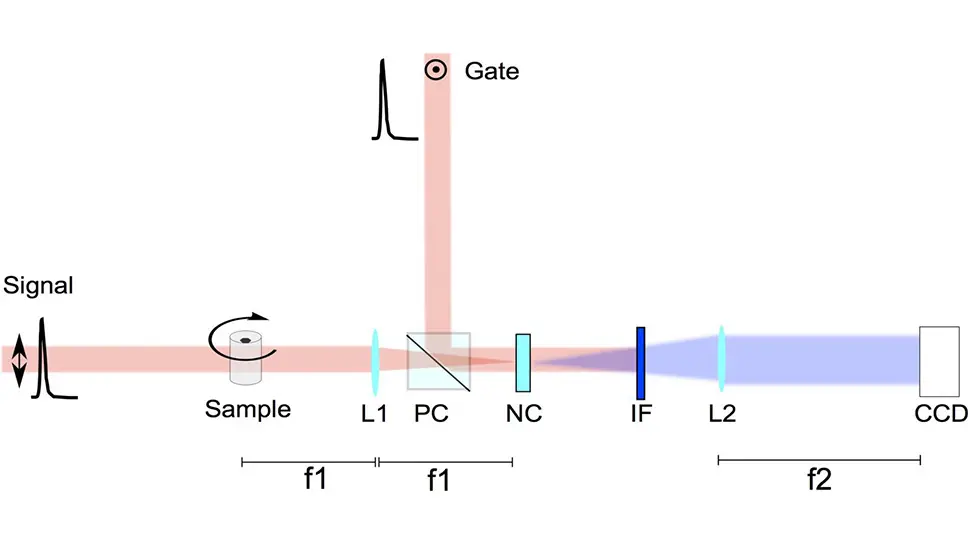
Furthermore we are developing an advanced version of OPT able to provide high resolution reconstructions of scattering samples, suitable for 3D imaging of larger biological specimens. The method consists in selecting the photons which propagate straight through the tissue (ballistic photons) by gating only a short temporal window of the transmitted light using a suitable non-linear optical process. We call this technique Time-Gated Optical Projection Tomography (TGOPT).
Here is the scheme of an optical setup for TGOPT
(fig.4 - Lenses (L1, L2); Polarizing Cube Splitter (PC); Non linear crystal (NC); Interference Filter (IF))
References:
- Andrea Bassi, Daniele Brida, Cosimo D’Andrea, Gianluca Valentini , Rinaldo Cubeddu, Sandro De Silvestri, Giulio Cerullo, "Time-gated optical projection tomography", Optics Letters 35, 2732-2734 (2010)
- Andrea Bassi, Luca Fieramonti, Cosimo D'Andrea, Marina Mione, Gianluca Valentini, "In-vivo label-free three-dimensional imaging of zebrafish vasculature with Optical Projection Tomography", Journal of Biomedical Optics (Letters) 16, 100502 (2011)
- Vadim Y. Soloviev, Andrea Bassi, Luca Fieramonti, Gianluca Valentini, Cosimo D’Andrea, and Simon R. Arridge, "Angularly selective mesoscopic tomography", Physical Review E 84(5), 051915, (2011)
- Luca Fieramonti, Andrea Bassi, Alesandro E. Foglia, Anna Pistocchi, Cosimo D'Andrea, Gianluca Valentini, Rinaldo Cubeddu, Sandro De Silvestri, Giulio Cerullo, Franco Cotelli "Time-Gated Optical Projection Tomography Allows Visualization of Adult Zebrafish Internal Structures", PLoS ONE 7(11), e50744 (2012)
- Alex Costa, Alessia Candeo, Luca Fieramonti, Gianluca Valentini, Andrea Bassi,
“Calcium Dynamics in Root Cells of Arabidopsis thaliana Visualized with Selective Plane Illumination Microscopy”, PLoS ONE, 8 (10), e75646, 1-11 (2013) - Luca Fieramonti, Efrem Foglia, Stefano Malavasi, Cosimo D'Andrea, Gianluca Valentini, Franco Cotelli, Andrea Bassi,
"Quantitative measurement of blood velocity in zebrafish with optical vector field tomography", Journal of Biophotonics, 8 (1-2), 52-57 (2015) - Andrea Bassi, Benjamin Schmid, Jan Huisken,
"Optical tomography complements light sheet microscopy for in toto imaging of zebrafish development", Development, 142(5), 1016-1020, (2015)
Research projects
NEXTSCREEN
Find out more