Start
01/04/2023
End
31/03/2027
Status
In progress
fastMOT
Website's ProjectStart
01/04/2023
End
31/03/2027
Status
In progress
fastMOT
Website's Project
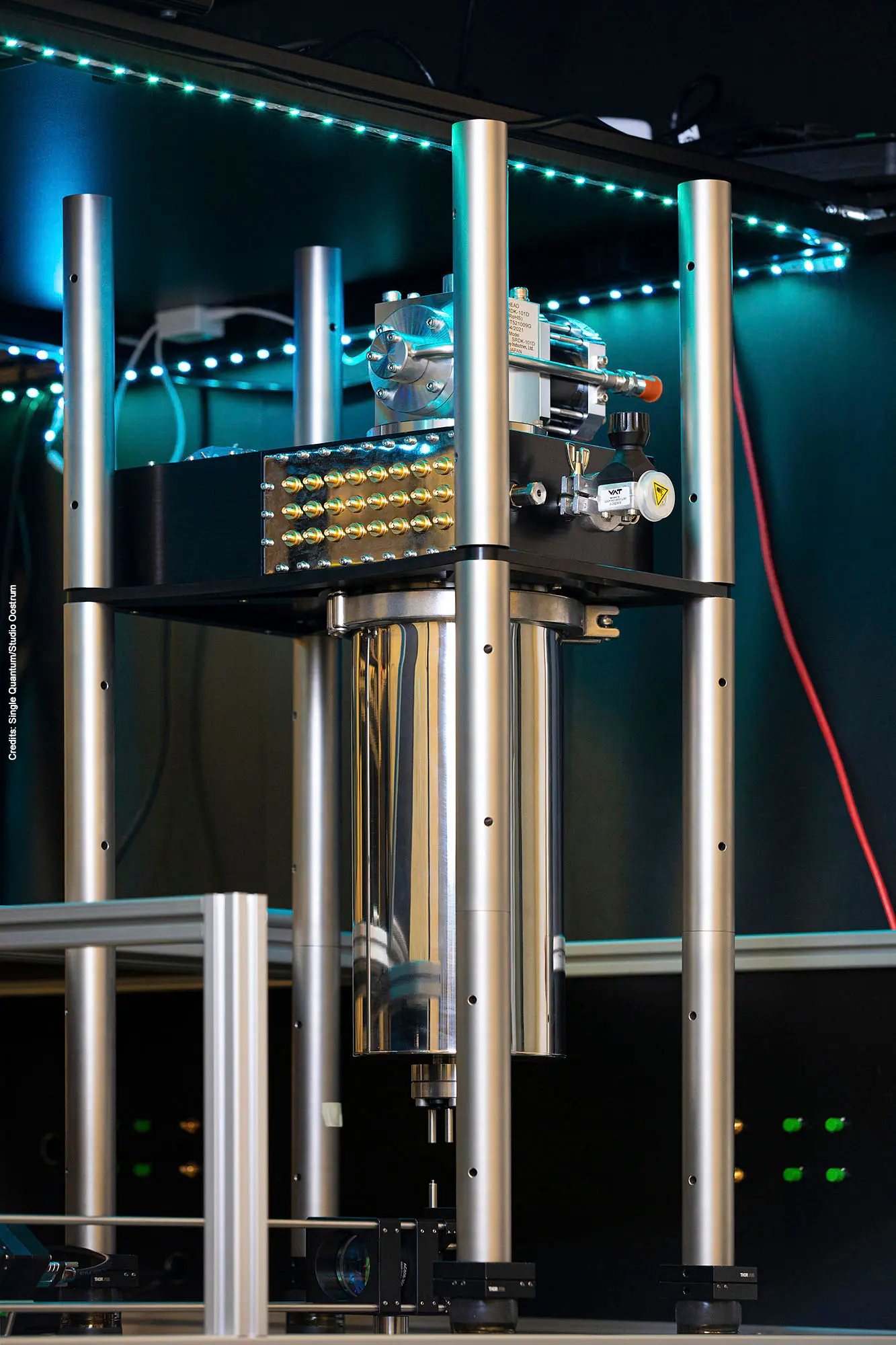
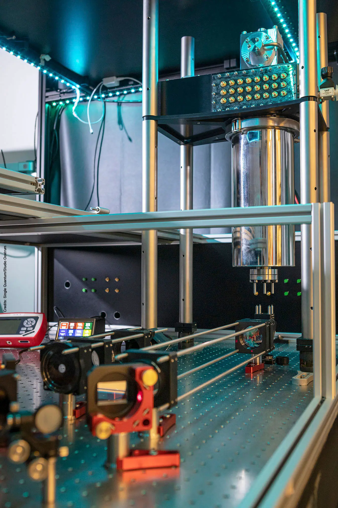
With its innovative fast gated, ultra-high quantum efficiency single-photon sensor, the fastMOT project will enable deep body imaging with diffuse optics. Implemented in the new Multifunctional Optical Tomograph, the light sensor will achieve a 100x improvement of signal-to-noise ratio compared to using existing light sensors.
Traditionally, organ monitoring and deep-body functional imaging are performed using ultrasound, X-ray (including CT), PET or MRI. However, these techniques allow only very limited measurements of functionality and are usually combined with exogenous and radioactive agents. To overcome this limitation, six partners, coordinated by the Dutch SME Single Quantum, have joined forces to develop an ultra-high performance light sensor in different imaging techniques to radically improve the performance of microscopy and imaging.
The novel sensor is based on superconducting nanowire single-photon detectors (SNSPDs), which have been shown to be ultra-fast and highly efficient. However, the active area and number of pixels have so far been limited to micrometre diameters and tens of pixels. The fastMOT consortium now aims at developing new techniques to overcome this limit and scale to 10,000 pixels and millimetre diameter. In addition, new strategies for performing time domain near infrared spectroscopy (TD-NIRS) and time domain speckle contrast optical spectroscopy (TD-SCOS) will be developed to optimally use this new light sensor with Monte-Carlo simulations. The new light sensor will be implemented in an optical tomograph and will achieve a 100x improvement of signal-to-noise ratio compared to using existing light sensors.
The role of the team from Physics Department (led by prof. Dalla Mora) is mainly focused on the development of the workstation (Multifunctional Optical Tomograph) which will be used for TD-NIRS and TD-SCOS, as well as for other applications.
Indeed, the proposed Multifunctional Optical Tomograph will make it possible to image deep organ and optical structures and monitor body functions such as oxygenation, haemodynamics, perfusion and metabolism. It also has the potential to significantly improve the accuracy of non-invasive breast cancer diagnosis, reducing the risk of false positive biopsies, with benefits for patients’ quality of life and improved sustainability for the healthcare systems.
Moreover, the Physics Department belongs to the Laserlab-Europe network (through CUSBO infrastructure) thus enabling the access to the developed workstation and its cutting-edge technologies to other scientists. This will increase the visibility of the developed workstation thus enabling ground breaking applications that will lead to new insights and a major economic boost in the years to come.
fastMOT (Fast gated superconducting nanowire camera for multi-functional optical tomograph) is funded by the EU’s HORIZON EUROPE programme (grant agreement 101099291) and by the UK Research and Innovation (UKRI) under the UK government’s Horizon Europe funding guarantee (grant number 10063660).
For more information see the fastMOT website or follow us on LinkedIn or X

01/02

02/02